The knee joint is one of the most complex joints in the body. It consists of bones, ligaments, and muscles. When any of these structures are injured you may have knee pain and difficulty in walking. You may hear a popping or snapping sensation at the time of the injury or you may feel like your knee is giving way. You may also have swelling, limping, and inability to move the knee.
If you have any of these symptoms, visit your doctor for an examination.
Your doctor will begin the knee examination by asking about your symptoms and history of any injury and then will perform a physical examination of your knee. Both medical history and physical examination are necessary for proper diagnosis and treatment plan.
For examination of your knee you may be asked to sit or lie down. Your doctor will examine both knees and compare your injured knee with the healthy knee.
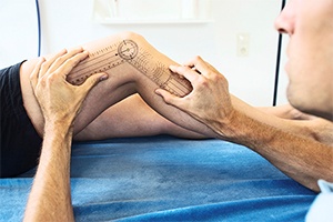
During the physical examination the following points will be noted:
Observation: Your doctor will assess your injured knee for redness, swelling, deformity, or any skin changes.

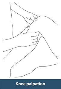
Palpation: Palpation is part of the examination in which your doctor feels your knee for temperature, swelling, tenderness, blood flow and altered sensation.
Range of motion: The doctor will evaluate your knee's range of motion by active and passive movements. In an active test, the patient is asked to move each joint through a full range of motion. In passive test, the joints are moved by the doctor. During this time, your doctor will listen for any popping, grinding or clicking sound in the joint.

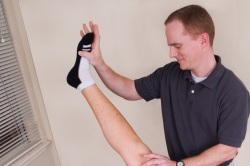
Active Test and Passive Test
The following tests are performed to evaluate the four ligaments of the knee:
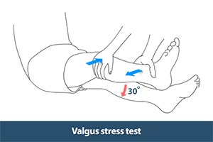
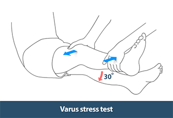
- Valgus and varus tests: Valgus and varus tests evaluate medial and lateral collateral ligaments. To perform these tests, your doctor will move your leg side-to-side by placing one hand on your knee joint and the other hand on your ankle.
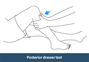
- Posterior drawer test: Posterior drawer test is done to evaluate the posterior cruciate ligament. To perform this test, the knee is placed at 90°angle and the foot is stabilized on the table. The doctor then places their hands around your knee and pushes the top of the knee with the thumb. Posterior subluxation of the tibia on the femur will result in patients with a PCL-deficient knee.
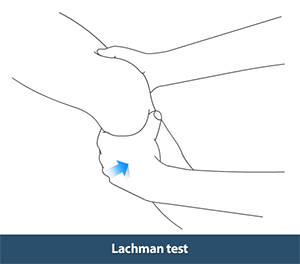
- Lachman test: This test is performed to diagnose an ACL tear. The knee is flexed slightly with the patient lying on their back. The doctor places one hand on the thigh while pulling the shin anteriorly to assess for any shifting of the shin bone as well and firmness or softness of the ligament.
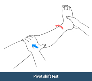
- Pivot shift test: Pivot shift test is a test used to evaluate an ACL. In this test, the leg is fully extended and your doctor applies a valgus stress to the knee with one hand and holds the ankle with other hand while internally rotating the tibia. If the ACL is torn the tibia will move ahead. Now slowly flex the knee while valgus and internal rotation is maintained. With an ACL tear, the knee will reduce with a clunk at around 30°of flexion. This test is usually performed before knee arthroscopy and after receiving anesthesia.
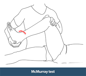
- McMurray test: This test is an important test for diagnosing meniscal tears. Your doctor holds your knee with one hand and the bottom of your foot with the other hand. Your leg is pushed up while turning the leg and pressing on the knee. Any pain or clicking sound during this test may suggest a meniscal tear.
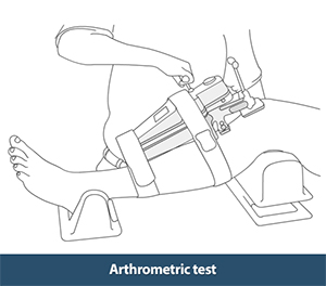
- Arthrometric test: Arthrometric test is usually helpful for patients with severe pain where it is difficult to perform examination. This test uses an instrument called an arthrometer; a device with two sensor pads and a pressure handle to measure the range of movement of your knee. The arthrometer is attached onto your lower leg with two sensor pads, one placed in contact with the patella (kneecap) and the other in contact with the tibial tubercle (a lump just below the knee cap). Your doctor then pulls or pushes on the pressure handle and measures the pressure. In addition to this, other tests may also be performed to evaluate the level of injury and to identify damage to other parts of the knee.
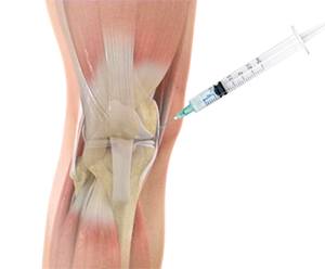
In some cases, if there is severe knee swelling your doctor may find it difficult to examine your knee. In these cases, a procedure called knee joint aspiration (arthrocentesis) is performed. Arthrocentesis or joint aspiration is a procedure in which joint fluid is removed from the joint to look for infection, inflammation, or bleeding. This test may also relieve pain and make the knee examination more comfortable.
The knee examination is a useful tool for your doctor to determine an appropriate diagnosis and to assist with developing a treatment plan. If you have any knee problems from a recent injury or from long lasting (chronic) symptoms, you may need to undergo a complete knee examination.
